Chapter: Human Nervous System and Sensory Organs : Basic Elements of the Nervous System
Blood Vessels
Blood Vessels
Cerebral blood vessels are of mesodermal origin. They grow during
development from the mesodermal coverings into the brain tissue. In
histological preparations, they are mostly surrounded by a narrow empty cleft (Virchow–Robin space, perivascular
space), an artifact caused by tissue shrinkage during histological preparation.
Arteries and arterioles are of the elastic
type, that is, their muscles are poorly developed and their contractility
is limited. The capillaries exhibit
a nonfenestrated closed endothelium
and a closed basal membrane. There
are nolymph vessels in the CNS.
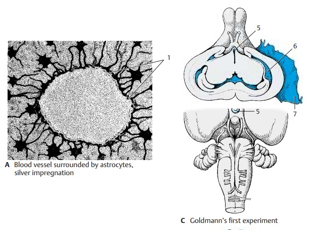
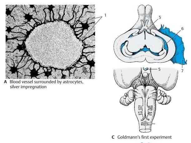
Astrocyte processes extend to the capillariesand widen into perivascular glial feet (AB1). In electron micrographs,
capillaries are completely covered by perivascular feet. The capillary wall
consists of endothelial cells (BE2) which overlap at their margins
like roof tiles and are joint together by zonulaeoccludentes
(tight junctions). The capillaryis enclosed by the basal lamina (BE3) and
the astrocyte covering (BE4). The latter can becompared to the glial limiting membrane; both structures
separate the ec-todermal tissue of the CNS from the adja-cent mesodermal
tissue.
The sealing of the brain tissue
from the rest of the body manifests itself in the blood–brain barrier, aselective
barrierfor numer-ous substances that are prevented from penetrating from
the bloodstream through the capillary wall into the brain tissue.
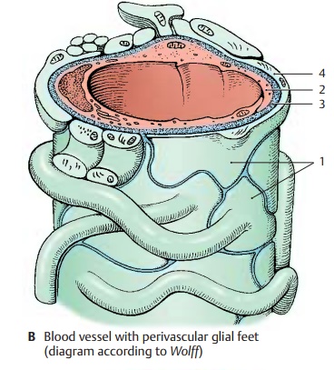
This barrier was first
demonstrated by Gold-mann’s experiments using trypan blue. If the dye is injected intravenously into
experi-mental animals (Goldmann’s first
experiment) (C), almost all
organs stain blue, but the brain and spinal cord remain unstained. Minor blue
staining is only found in the graytubercle
(C5), the postremal area, and the
spinal ganglia. The choroid plexus (C6) andthe dura (C7) show a
distinct blue staining. The same pattern is observed in cases of jaundice in
humans; the bile pigment stains all organs yellow, only the CNS remains
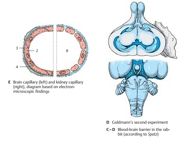
un-stained. If the dye is injected into the spaceof the cerebrospinal fluid (Goldmann’s secondexperiment) (D), brain and spinal cord arediffusely
stained on the surface, while the rest of the body remains unstained. Thus,
there exists a barrier between the CSF and the blood but not between the CSF
and the CNS. We therefore distinguish between a blood–brain barrier and a blood–cere-brospinal fluid barrier. The
two barriersbehave in different ways.
The site of the blood–brain
barrier is the capillary endothelium (E) (see also vol. 2);in the brain, it
forms a closed wall without fenestration. By contrast, the capillary walls of
many other organs (liver, kidney [E8])
ex-hibit prominent fenestration which allows for extensive metabolite exchange.
The bar-rier effect has been demonstrated for numerous substances in studies
using iso-topes. The barrier may result in a complete blockade or in a delay of
penetration. Whether or not drugs can penetrate this barrier has major
practical implications.
Related Topics