Chapter: Essentials of Anatomy and Physiology: Blood Vessels and Circulation
Blood Vessels of the Systemic Circulation: Arteries
BLOOD VESSELS OF THE SYSTEMIC CIRCULATION: ARTERIES
The systemic circulation is the system of blood vessels that carries blood from the left ventricle of the heart to the tissues of the body and back to the right atrium. Oxygen-rich blood from the pulmo-nary veins passes from the left atrium into the left ventricle and from the left ventricle into the aorta. Arteries distribute blood from the aorta to all portions of the body (figure 13.6).

Aorta
All arteries of the systemic circulation branch directly or indi-rectly from the aorta (ā -ō r′ t̆a ). The aorta is usually considered in three parts—the ascending aorta, the aortic arch, and the descend-ing aorta; the last segment is further divided into the thoracic aorta and the abdominal aorta (figure 13.7a).
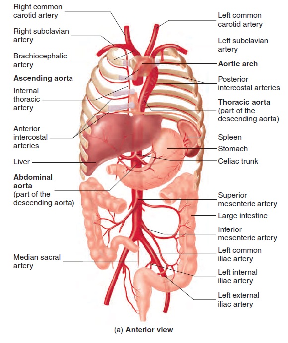
The ascending aorta is the part of the aorta that passes supe-riorly from the left ventricle. The right and left coronary arteries arise from the base of the ascending aorta and supply blood to the heart .
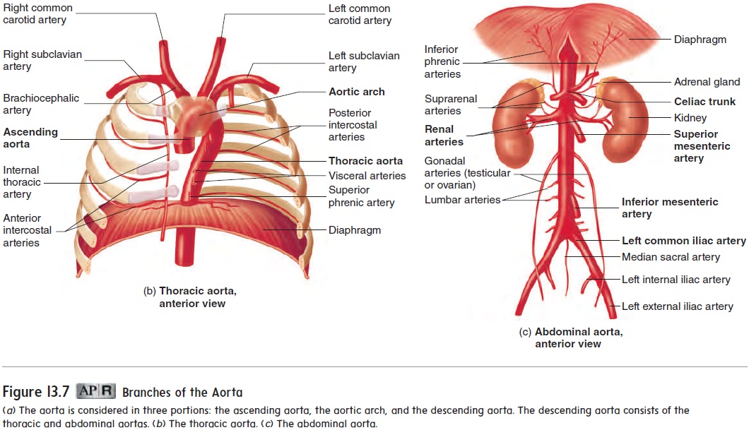
The aorta arches posteriorly and to the left as the aortic arch. Three major arteries, which carry blood to the head and upper limbs, originate from the aortic arch: the brachiocephalic artery, the left common carotid artery, and the left subclavian artery (figure 13.7b).
The descending aorta is the longest part of the aorta. It extends through the thorax and abdomen to the upper margin of the pelvis. The part of the descending aorta that extends through the thorax tothe diaphragm is called the thoracic (thō -ras′ ik) aorta. The part of the descending aorta that extends from the diaphragm to the point at which it divides into the two common iliac (il′ ē -ak) arteries is called theabdominal (ab-dom′ i-n̆a l) aorta (figure 13.7a,c).
An arterial aneurysm (an′ ū -rizm; a dilation) is a localized dilation of an artery that usually develops in response to trauma or a congenital (existing at birth) weakness of the artery wall. Rupture of a large aneurysm in the aorta is almost always fatal, and rupture of an aneurysm in an artery of the brain causes massive damage to brain tissue and even death. If aneurysms are discovered, they can sometimes be surgically corrected. For example, large aortic aneurysms that leak blood slowly can often be repaired.
Arteries of the head and neck
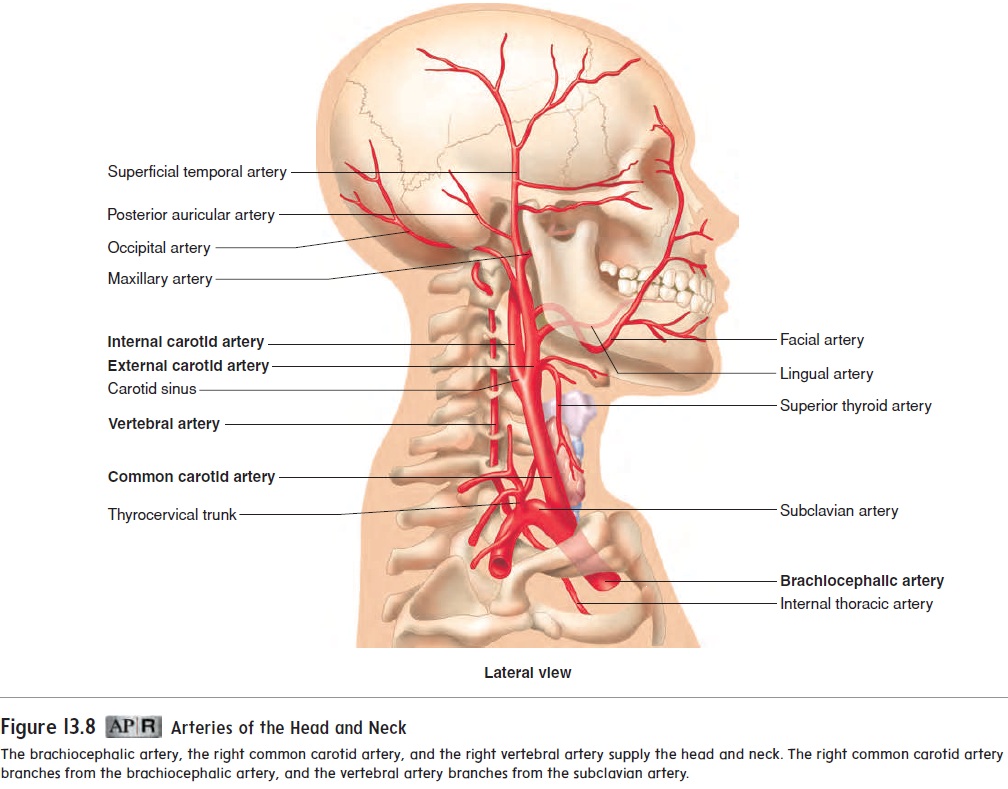
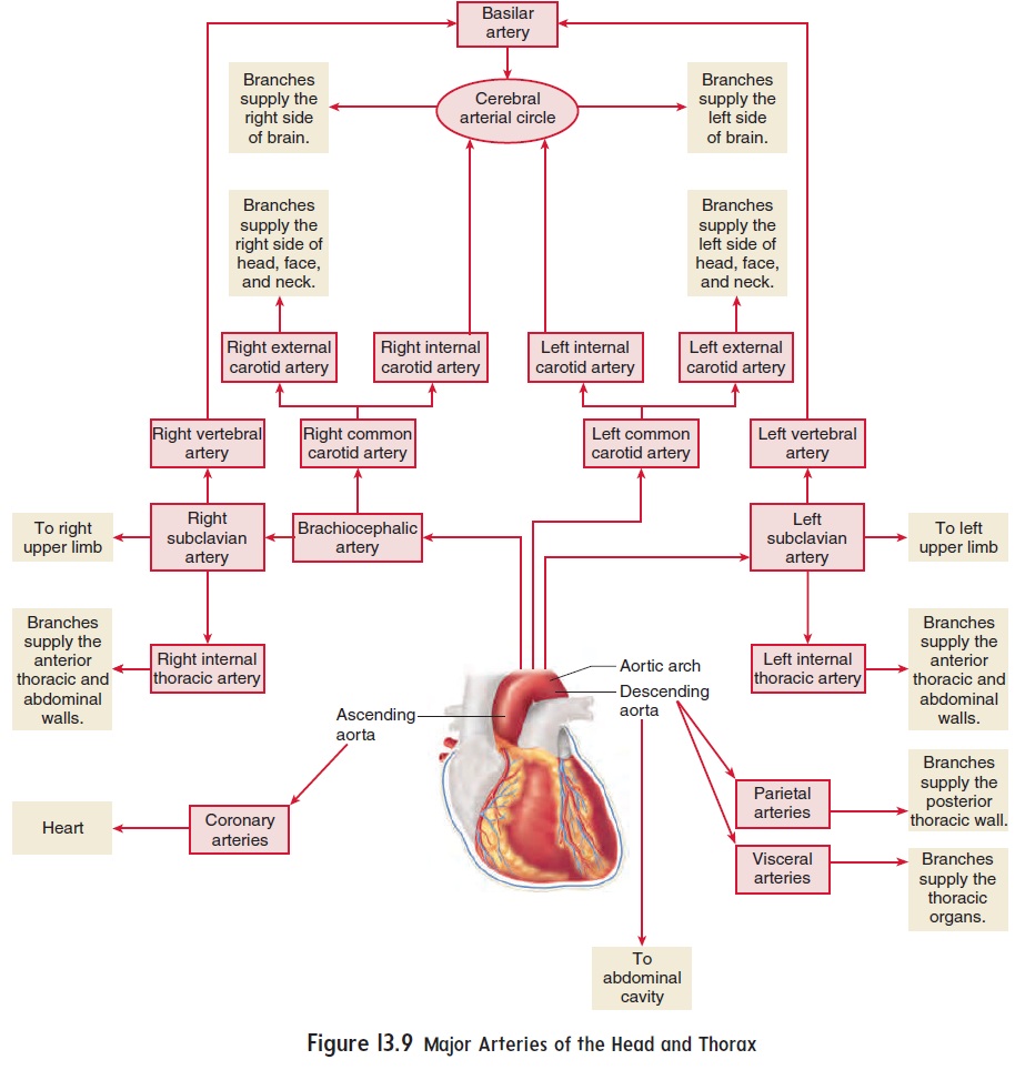
Figure 13.8 depicts the arteries of the head and neck, and figure 13.9 shows how these arteries connect to each other and to the arteries of the thorax. It may help you to refer to figure 13.9 as you read the following discussion. The first vessel to branch from the aor-tic arch is the brachiocephalic (br̄a ′ kē - ō -se-fal′ ik; vessel to the arm and head) artery. This short artery branches at the level of the clavicle to form the right common carotid (ka-rot′ id) artery, which transports blood to the right side of the head and neck, and the right subclavian (sŭb-kl̄a′vē-an; beneath the clavicle) artery, which transports blood to the right upper limb (see figures 13.7b and 13.8).
There is no brachiocephalic artery on the left side of the body. Instead, both the left common carotid and the left subclavian arter-ies branch directly off the aortic arch (see figures 13.6, 13.7a,b, and 13.9). They are the second and third branches of the aortic arch. The left common carotid artery transports blood to the left side of the head and neck, and the left subclavian artery transports blood to the left upper limb.
The common carotid arteries extend superiorly along each side of the neck to the angle of the mandible, where they branch into internal and external carotid arteries. The base of each internal carotid artery is slightly dilated to form a carotid sinus, which contains structures important in monitoring blood pres-sure (baroreceptors). The external carotid arteries have several branches that supply the structures of the neck, face, nose, and mouth. The internal carotid arteries pass through the carotid canals and contribute to the cerebral arterial circle (circle of Willis) at the base of the brain (figure 13.10). The vessels that supply blood to most of the brain branch from the cerebral arterial circle.
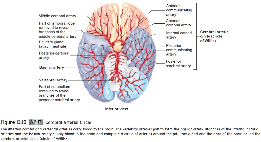
Some of the blood to the brain is supplied by the vertebral (ver′tĕ-brăl) arteries, which branch from the subclavian arteries and pass to the head through the transverse foramina of the cervical vertebrae (see figure 13.8). The vertebral arteries then pass into the cranial cavity through the foramen magnum. Branches of the vertebral arteries supply blood to the spinal cord, as well as to the vertebrae, muscles, and ligaments in the neck.
Within the cranial cavity, the vertebral arteries unite to form a single basilar (bas′i-lăr; relating to the base of the brain) artery located along the anterior, inferior surface of the brainstem (fig-ure 13.10). The basilar artery gives off branches that supply blood to the pons, cerebellum, and midbrain. It also forms right and left branches that contribute to the cerebral arterial circle. Most of the blood supply to the brain is through the internal carotid arteries; however, not enough blood is supplied to the brain to maintain life if either the vertebral arteries or the carotid arteries are blocked.
Arteries of the Upper Limbs
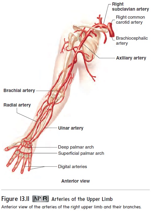
The arteries of the upper limbs are named differently as they pass into different body regions, even though no major branching occurs. The subclavian artery, located deep to the clavicle, becomes the axillary(ak′sil-ār-ē) artery in the axilla (armpit). The brachial (brā′kē-ăl) artery, located in the arm, is a continuation of the axillary artery (figure 13.11). Blood pressure measurements are normally taken from the brachial artery. The brachial artery branches at the elbow to form the ulnar (ul′năr) artery and the radial (rā′dē-ăl) artery, which supply blood to the forearm andhand. The radial artery is the one most commonly used for taking a pulse. The pulse can be detected easily on the thumb (radial) side of the anterior surface of the wrist
Thoracic Aorta And Its Branches
The branches of the thoracic aorta can be divided into two groups: The visceral (vis′ er-̆a l) arteries supply the thoracic organs, and the parietal (p̆a -r ′ ̆e -t̆a l) arteries supply the thoracic wall. The visceral branches of the thoracic aorta supply the esophagus, the trachea, the parietal pericardium, and part of the lung. The major parietal arteries are the posterior intercostal (in-ter-kos′ t̆a l) arteries, which arise from the thoracic aorta and extend between the ribs (see figure 13.7a,b). They supply the intercostal muscles, the vertebrae, the spinal cord, and the deep muscles of the back. The superior phrenic (fren′ ik; diaphragm) arteries supply the diaphragm.
The internal thoracic arteries are branches of the subclavian arteries. They descend along the internal surface of the anterior thoracic wall and give rise to branches called the anterior intercostalarteries,which extend between the ribs to supply the anterior chestwall (see figure 13.7a,b).
Abdominal Aorta and its Branches
The branches of the abdominal aorta, like those of the thoracic aorta, can be divided into visceral and parietal groups. The visceral arter-ies are divided into paired and unpaired branches. There are three major unpaired branches: the celiac (sē ′ lē -ak; belly) trunk, the superior mesenteric (mez-en-ter′ik) artery, and the inferior mesenteric artery. The celiac trunk supplies blood to the stomach,pancreas, spleen, upper duodenum, and liver.
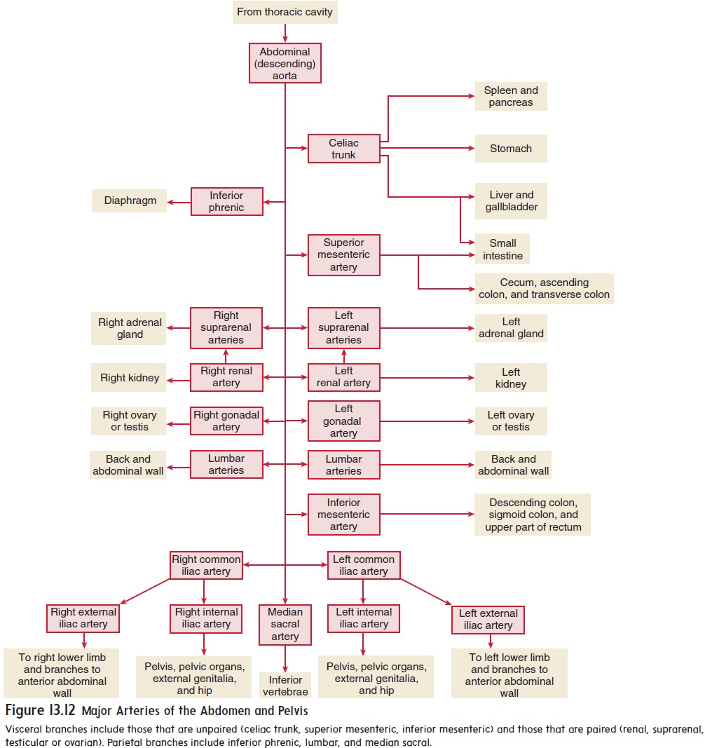
The superior mesenteric artery supplies blood to the small intestine and the upper portion of the large intestine, and the inferior mesenteric artery supplies blood to the remainder of the large intestine (figure 13.12).
There are three paired visceral branches of the abdominal aorta. The renal (rē′ nal; kidney) arteries supply the kidneys, and the suprarenal (sū′ pră-rē ′ năl; superior to the kidney) arteries supply the adrenal glands. The testicular arteries supply the tes-tes in males; the ovarian arteries supply the ovaries in females.
The parietal branches of the abdominal aorta supply the diaphragm and abdominal wall. The inferior phrenic arteries supply the diaphragm; the lumbar arteries supply the lumbar vertebrae and back muscles; and the median sacral artery sup-plies the inferior vertebrae.
Arteries of the Pelvis
The abdominal aorta divides at the level of the fifth lumbar ver-tebra into two common iliac arteries. Each common iliac artery divides to form an external iliac artery, which enters a lower limb, and aninternal iliac artery, which supplies the pelvic area (see figure 13.7c). Visceral branches of the internal iliac artery supply organs such as the urinary bladder, rectum, uterus, and vagina. Parietal branches supply blood to the walls and floor of the pelvis; the lumbar, gluteal, and proximal thigh muscles; and the external genitalia.
Arteries of the lower limbs
Like the arteries of the upper limbs, the arteries of the lower limbs are named differently as they pass into different body regions, even though there are no major branches.
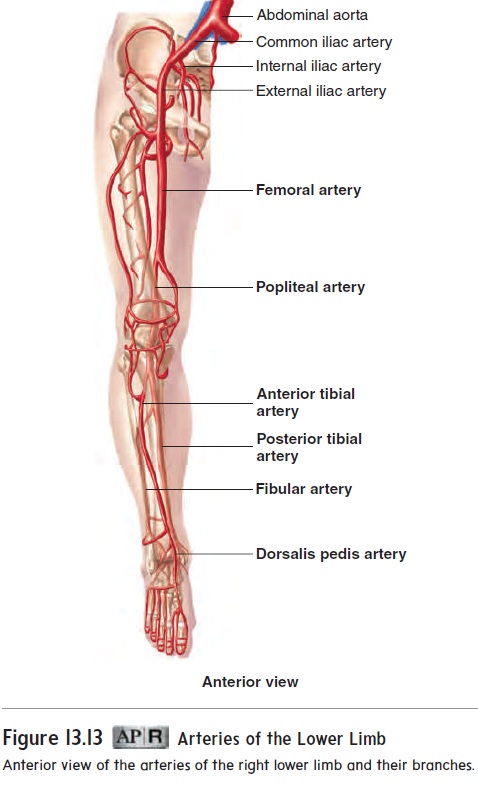
The external iliac artery in the pelvis becomes the femoral (fem′ ŏ-răl) artery in the thigh, and it becomes the popliteal (pop-lit′ ē-ăl) artery in the popliteal space, which is the posterior region of the knee. The popliteal artery branches slightly inferior to the knee to give off the anterior tibialartery and the posterior tibial artery, both of which give rise toarteries that supply blood to the leg and foot (figure 13.13). The anterior tibial artery becomes the dorsalis pedis (dōr-sāl′ lis pē′ dis pes, foot)arteryat the ankle. The posterior tibial artery gives riseto the fibular artery, or peroneal artery, which supplies the lateral leg and foot.
The femoral triangle is located in the superior and medial area of the thigh. Its margins are formed by the inguinal ligament, the medial margin of the sartorius muscle, and the lateral margin of the adductor longus muscle (see figures 7.14a and 7.26a). Passing
through the femoral triangle are the femoral artery, vein, and nerve. A pulse in the femoral artery can be detected in the area of the femoral triangle. This area is also susceptible to serious traumatic injuries that result in hemorrhage and nerve damage. In addition, pressure applied to this area can help prevent bleeding from wounds in more distal areas of the lower limb. The femoral triangle is an important access point for certain medical proce-dures as well.
Related Topics