Chapter: Human Nervous System and Sensory Organs : Spinal Cord and Spinal Nerves
Blood Vessels of the Spinal Cord
Blood Vessels of the Spinal Cord
The spinal cord is supplied with
blood from two sources, the vertebral
arteries and the segmental arteries (intercostal arteries and lumbar arteries).
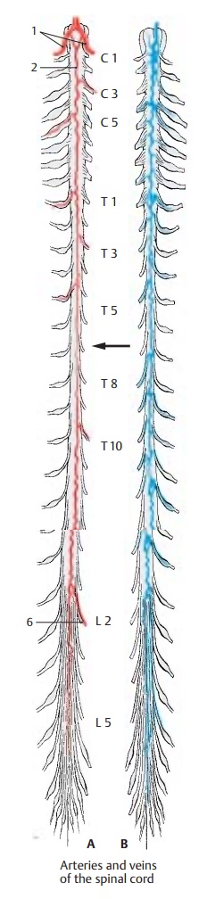
Vertebral arteries (A1).Before they unite,they give off two thin posterior spinal arter-ies that form a
network of small arteriesalong the posterior surface of the spinal cord. At the
level of the pyramidal decussa-tion, two additional branches of the verte-bral
arteries join to form the anterior
spinalartery (AD2) which runs
along the anteriorsurface of the spinal cord at the entrance to the anterior
sulcus.
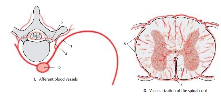
Segmental arteries (C3).Their posteriorbranches (C4) and the vertebral arteries give off spinal branches (C5)
which enter through the intervertebral foramina and divide at the spinal roots
into dorsal and ventral branches to supply the spinal roots and the spinal
meninges. Of the 31 spinal arteries, only 8 to 10 extend to the spinal cord and
contribute to its blood supply. The levels at which the radicular arteries
approach the spinal cord vary, and so do the sizes of the vessels. The largest
vessel approaches the spinal cord at the level of the lumbar en-largement
between T12 and L3 (large radicu-lar
artery) (A6).
The anterior spinal artery is widest at the level of the cervical and
lumbar enlargements. Its diameter is much reduced in the mid-thoracic region of
the spinal cord. As this re-gion is also the border area between two supplying
radicular arteries, this segment of the spinal cord is especially at risk in
case of circulatory problems (A,
arrow). Depending on the variation of the radicular arteries, this may also
apply to other segments of the spinal cord.
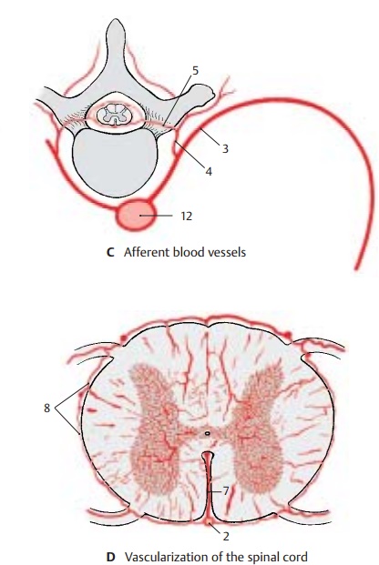
The anterior spinal artery gives
off numer-ous small arteries into the anterior sulcus, the sulcocommissural arteries (D7).
In the cer-vical and thoracic spinal cords, they turn al-ternately to the left
and right halves of the spinal cord; in the lumbar and sacral spinal cords,
they divide into two branches. In ad-dition, anastomoses arise between the
ante-rior and posterior spinal arteries, so that the spinal cord is surrounded
by a vascular ring (vasocorona) (D8) from where vessels radiate into the
white matter. Injection of tracers revealed that the gray matter is much more
vascularized than the white matter (D).
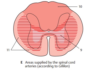
Areas of blood supply (E).The anterior spi-nal artery supplies the anterior horns,
the bases of the posterior horns, and the largest part of the anterior lateral
funiculi (E9). The posterior
funiculi and the remaining parts of the posterior horns are supplied by the posterior spinal arteries (E10). The marginalzone of the anterior
lateral funiculus is sup-plied by the plexus of the vasocorona (E11).
The spinal veins (B) form a
network in which one anterior spinal vein
and two pos-terior spinal veins stand
out. The efferentveins run along the spinal roots and open into the epidural venous plexus (see vol. 2). The
spinal veins lack valves prior to their penetration through the dura.
C12 Aorta.
Related Topics