Chapter: Medical Physiology: Cardiac Arrhythmias and Their Electrocardiographic Interpretation
Atrial Paroxysmal Tachycardia
Atrial Paroxysmal Tachycardia
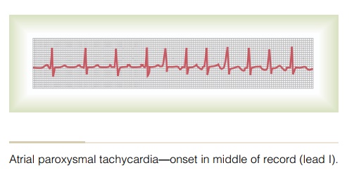
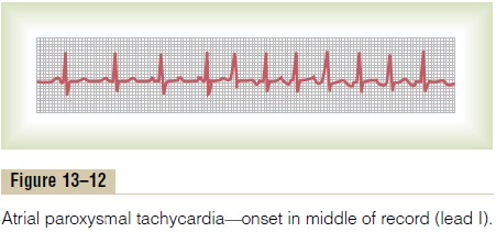
Figure 13–12 demonstrates in the middle of the record a sudden increase in the heart rate from about 95 to about 150 beats per minute. On close study of the elec-trocardiogram during the rapid heartbeat, an inverted P wave is seen before each QRS-T complex, and this P wave is partially superimposed onto the normal T wave of the preceding beat. This indicates that the origin of this paroxysmal tachycardia is in the atrium, but because the P wave is abnormal in shape, the origin is not near the sinus node.
A-V Nodal Paroxysmal Tachycardia. Paroxysmal tachycardiaoften results from an aberrant rhythm that involves the A-V node. This usually causes almost normal QRS-T complexes but totally missing or obscured P waves.
Atrial or A-V nodal paroxysmal tachycardia, both of which are called supraventricular tachycardias, usually occurs in young, otherwise healthy people, and they gen-erally grow out of the predisposition to tachycardia after adolescence. In general, supraventricular tachy-cardia frightens a person tremendously and may cause weakness during the paroxysm, but only seldom does permanent harm come from the attack.
Related Topics