Chapter: Human Nervous System and Sensory Organs : The Eye
Anterior Part of the Eye
Anterior Part of the Eye
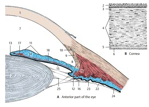
Cornea (A, B)
The cornea (A1, B) is positioned like a watch-glass on the eyeball. Because of its marked curvature, it has the effect of a focusing lens. Its anterior surface is formed by a multi-layered, nonkeratinizing squamous epi-thelium (B2) that rests upon a thick base-ment membrane, called the anterior limitinglamina (Bowman’s membrane) (B3). Belowthis lies the stroma of the cornea (substantiapropria) (B4); its straight collagen fibers form lamellae that lie parallel to the surface of the cornea. At the posterior surface lies another basement membrane, the posteriorlimiting lamina (Descemet’s membrane)(B5) and a single-layered endothelium (B6). The cornea contains unmyelinated nerve fibers but no blood vessels. Its transparency is due to a specific fluid content and the state of swelling of its components. Any change in the turgor causes turbidity of the cornea.

Anterior Chamber of the Eye (A)
The anterior chamber of the eye (A7) con-tains the aqueous humor generated by the blood vessels of the iris. The wall of the iridocorneal angle (A8) consists of looseconnective-tissue strands (reticulumtrabeculare, orpectinate ligament) (A9); theaqueous humor filters through the spaces between the fibers into the venous sinus ofthe sclera, or Schlemm’s canal (A10), andpasses into the bloodstream.
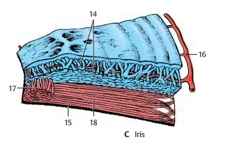
Iris (A, C)
The iris (A11) forms an aperture in front of the lens. It is attached to the ciliary body at the root of the iris (A12) and extends to the pupillary margin (A13). The iris consists oftwo layers, namely, the mesodermal stroma (AC14) and the ectodermal posterior aspect of the iris, known as the iridial part of theretina (AC15). The stroma is made up of con-nective-tissue strands and is pigmented. A high pigment content results in brown eyes, while a low pigment content gives the eyes a green or blue appearance. Numerousblood vessels branch radially off the greaterarterial circle of the iris (AC16). The ectoder-mal part of the iris, a derivative of the optic cup, gives rise to two smooth muscles, namely, the sphincter muscle of thepupil (AC17) and the thin dilator muscle of the pupil (AC18).
Ciliary Body (A, D)
The lens is suspended at the circular ciliarybody (A19, D). The muscles of the ciliarybody control the curvature of the lens and, hence, the visual acuity for close and distant vision. It consists of a radially folded surface, the ciliary disk (D20), from where approximately 80 ciliaryprocesses (D21) protrude (ciliary crown). Theanterior part of the disk is occupied by the ciliary muscle; its muscle fibers, the merid-ional fibers (A22), span between the oraser-rata and the reticulum trabeculare as well as Descemet’s membrane. From here, radial muscle fibers extend inward and bend to take a circular course (circular fibers) (A23). The posterior surface of the ciliary body is covered by the ciliary part of the retina(A24). Delicate fibers, the zonular fibers (AD25), extend from the ciliary body to the lens and form the ciliary zone (D26). Many fibers originate from the area of the oraser-rata and extend to the anterior surface of the lens. They cross the shorter fibers aris-ing from the ciliary processes and ending at the posterior surface of the lens.
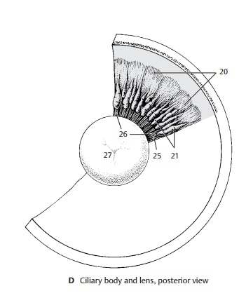
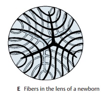
Lens of the Eye (A, D, E)
The biconvex lens (AD27) is made up by elongated epithelial cells, the lens fibers, which show a lamellar arrangement. The ends of the fibers push against each other at the anterior and posterior surfaces of the lens; in the newborn, they form a three-pointed star (E) at their border. The lens fibers are continuously being formed throughout life.
Related Topics