Chapter: Obstetrics and Gynecology: Embryology and Anatomy
Anomalies of the Female Reproductive System
ANOMALIES OF THE FEMALE REPRODUCTIVE SYSTEM
Anatomic anomalies are infrequent
and arise from defects during embryologic development. Ovarian dysgenesis or congenital absence is rare except in cases of
chro-mosomally abnormalities. In Turner syndrome (45XO), there are streaks of
abnormal ovarian tissues in the pelvis. In the anatomically female patient with
a male chromosome compliment (46XY), the gonads only partially descend and can
usually be found in the pelvis or even in the inguinal canal.
Much more
common are müllerian (paramesonephric) abnormalities, most of which stem from
incomplete or anom-alous fusion of the müllerian ducts. Absence
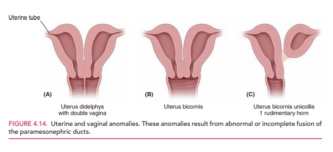
of the uterusoccurs when the müllerian ducts degenerate, a condition called müllerian agenesis (Fig. 4.14). This
condition is associated with vaginal anomalies (such as absence of the vagina),
because vaginal development is stimulated by the developing uterovaginal
primordium. Since the vulva and the external portion of the vagina develop from
the invagination of the urogenital sinus, the exter-nal genitalia can appear
normal in these women. A dou-ble uterus (uterus
didelphys) occurs when the inferior parts of the müllerian ducts do not
fuse; this condition may be associated with a double or a single vagina. A bicornuate uterus results when lack of
fusion is limitedto the superior portion of the uterine body. If one of the
ducts is poorly developed and fusion with the other duct does not occur, the
result is a bicornuate uterus with a rudimentary horn. This horn may or may not commu-nicate with the uterine
cavity.

The mesonephric ducts normally
degenerate in the female embryo during development of the reproductive tract.
However, remnants of the mesonephric ducts can persist, which can manifest as
Gartner cysts (Fig. 4.15).
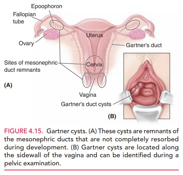
These cysts are located along the
vaginal wall or within the broad ligament of the uterus.
Since the paramesonephric system
develops alongside the renal system, frequently when one system is abnormally
formed, an abnormality in the other is frequently present. For example, in a
woman with renal agenesis on one side, an abnormal fallopian tube is often
found. Conversely, despite the functional connection between the ovaries and
fallopian tubes, a lack of one does not indicate a probable lack of the other.
Related Topics