Chapter: Obstetric and Gynecological Nursing : Anatomy of Female Pelvis and the Fetal Skull
Anatomy of the female external genitalia

Anatomy of the female external
genitalia
The vulva
This term applies to the external female genital organs. It consists of
the following structures. The mons pubis
ormons veneris - is a pad of fat lying over the Symphysispubis. It is
covered with pubic hair from the time of puberty.
The labia majora (greater lips)
The labia minora (lesser lips) anteriorly encloses clitoris
andposteriorlny forms furchette.
The clitoris is a small rounded organ of
erectile tissue at theforwarded junction of the labia minora.
The vestibule is the flattend, smooth surface
in side the labiaThe vaginal orifice
Bartholin's glands (volvovaginal
glands) are
located justlateral to the vaginal opening on the sides.
The furchette is ridge of tissue formed by the
posterior joiningof the two labia minora and the labia majora.
The vulval blood supply comes mainly from the
pudendalarteries and apportion of the inferior rectus aretery. The blood drains
through the pundendal veins.
Lymphatic drainage - inpuinal glands
Nerve supply - branch of pudendal nerve
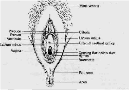
Figure 6 . Female external genitalia
The vagina
Position –
is a canal running from the vestibule to the cervix.
Relations:-
A knowledge of the relation of the vagina is essential for the accurate
examination of the pregnant woman and her safe delivery.It is found infront of
the rectum and behind the bladder and urthrea.
Structure
the posterior wall is longer than the antrerior
the vaginal walls are pink in appearance and thrown into small folds known as
rugae. These allow the vaginal wall to stretch during intercourse and child
birth.
Layers
squamins epithelium, vascular connective tissue,
weak inner coat of circular fibers and stronger outer coat of longitudinal
fibers. Pelvic fascia surrounds the vagina forming a layer of connective
tissue.
Contents
the vaginal fluid is strongly acidic (PH 4.5)
Blood supply
from braches of the internal iliac artery and
drains through corresponding Veins.
Lymphatic drainage
via the inguinal, the internal iliac and the
sacral glands drains the lymphatic fluid.
Related Topics