Chapter: Essentials of Anatomy and Physiology: Digestive System
Anatomy of the Small intestine
Anatomy of the Small intestine
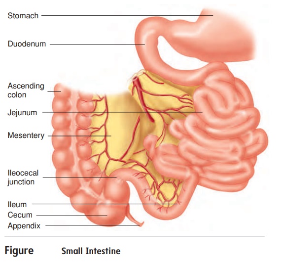
The small intestine is about 6 meters (m) long and consists of three parts: the duodenum, the jejunum, and the ileum (figure 16.13). The duodenum (doo-od′̆e-n̆um, doo-̄o-d̆e′n̆um) is about 25 cm long(the term duodenum means 12, suggesting that it is 12 in. long). The jejunum (j̆e -joo′ n̆u m) is about 2.5 m long and makes up two-fifths of the total length of the small intestine. The ileum (il′ ̄e -̆u m) is about 3.5 m long and makes up three-fifths of the small intestine.
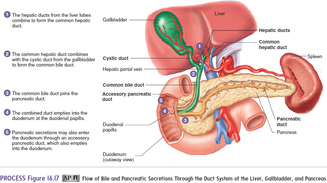
The duodenum nearly completes a 180-degree arc as it curves within the abdominal cavity. Part of the pancreas lies within this arc. The common bile duct from the liver and the pancreatic duct from the pancreas join and empty into the duo-denum (see figure 16.17).
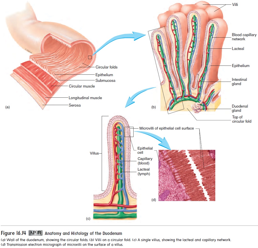
The small intestine is the major site of digestion and absorp-tion of food, which are accomplished due to the presence of a large surface area. The small intestine has three modifications that increase its surface area about 600-fold: circular folds, villi, and microvilli. The mucosa and submucosa form a series of circularfolds that run perpendicular to the long axis of the digestive tract(figure 16.14a). Tiny, fingerlike projections of the mucosa form numerous villi (vil′ i; sing. villus), which are 0.5–1.5 mm long (figure 16.14b).
(mı̄′ krō-vil′ ı̄) (figure 16.14c,d). Each villus is covered by simple columnar epithelium. Within the lMost of the cells composing the surface of the villi have numerous cytoplasmic extensions, called microvilli oose connective tissue core of each villus are a blood capillary network and a lymphatic capil-lary called a lacteal (lak′ tē-ăl; resembling milk) (figure 16.14c). The blood capillary network and the lacteal are very important in transporting absorbed nutrients.
The mucosa of the small intestine is simple columnar epi-thelium with four major cell types: (1) absorptive cells, which have microvilli, produce digestive enzymes, and absorb digest-ed food; (2) goblet cells, which produce a protective mucus;(3) granular cells, which may help protect the intestinal epi-thelium from bacteria; and (4) endocrine cells, which produce regulatory hormones.
The epithelial cells are located within tubular glands of the mucosa, called intestinal glands or crypts of Lieberkühn, at the base of the villi. Granular and endocrine cells are located in the bottom of the glands. The submucosa of the duodenum contains mucous glands, called duodenal glands, which open into the base of the intestinal glands.
The duodenum, jejunum, and ileum are similar in structure. However, progressing from the duodenum through the ileum, there are gradual decreases in the diameter of the small intestine, in the thickness of the intestinal wall, in the number of circular folds, and in the number of villi. Lymphatic nodules are common along the entire length of the digestive tract, and clusters of lym-phatic nodules, called Peyer patches, are numerous in the ileum. These lymphatic tissues help protect the intestinal tract from harmful pathogens.
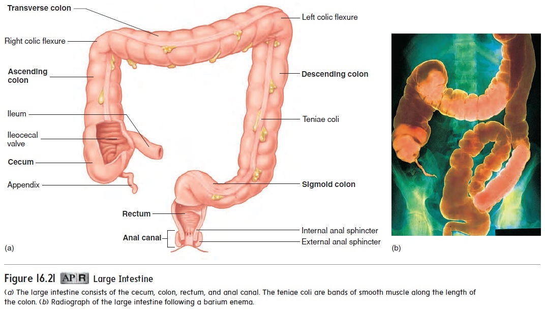
The site where the ileum connects to the large intestine is called the ileocecal (il′ ē-ō-sē′ kăl) junction. It has a ring of smooth muscle, the ileocecal sphincter, and an ileocecal valve (see figure 16.21a), which allow the intestinal contents to move from the ileum to the large intestine, but not in the opposite direction.
Related Topics