Chapter: Medical Surgical Nursing: Assessment and Management of Patients With Hearing and Balance Disorders
Anatomy of the Middle Ear
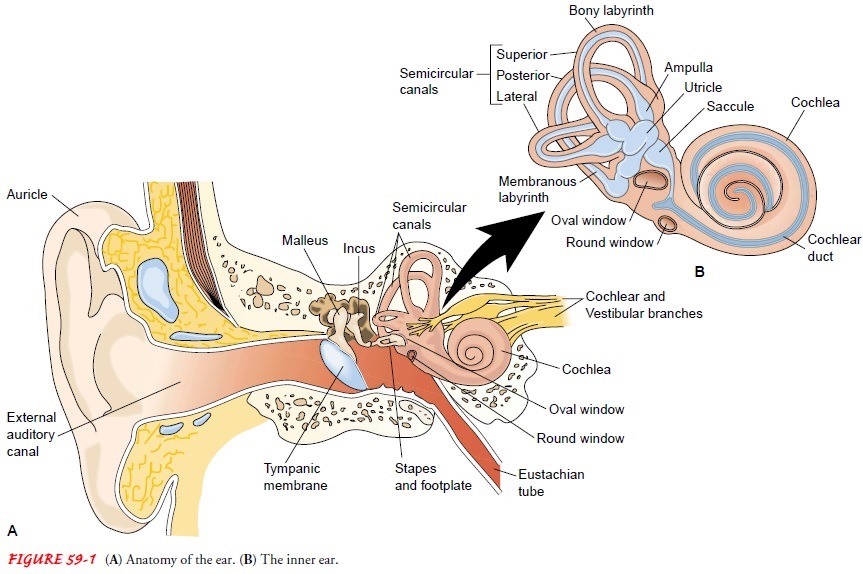
ANATOMY OF THE MIDDLE EAR
The
middle ear, an air-filled cavity, includes the tympanic mem-brane laterally and
the otic capsule medially. The middle ear cleft lies between the two. The
middle ear is connected by the eusta-chian tube to the nasopharynx and is
continuous with air-filled cells in the adjacent mastoid portion of the
temporal bone.
The
eustachian tube, which is approximately 1 mm wide and 35 mm long, connects the
middle ear to the nasopharynx. Nor-mally, the eustachian tube is closed, but it
opens by action of the tensor veli palatini muscle when performing a Valsalva
ma-neuver or when yawning or swallowing. The tube serves as a drainage channel
for normal and abnormal secretions of the middle ear and equalizes pressure in
the middle ear with that of the atmosphere.
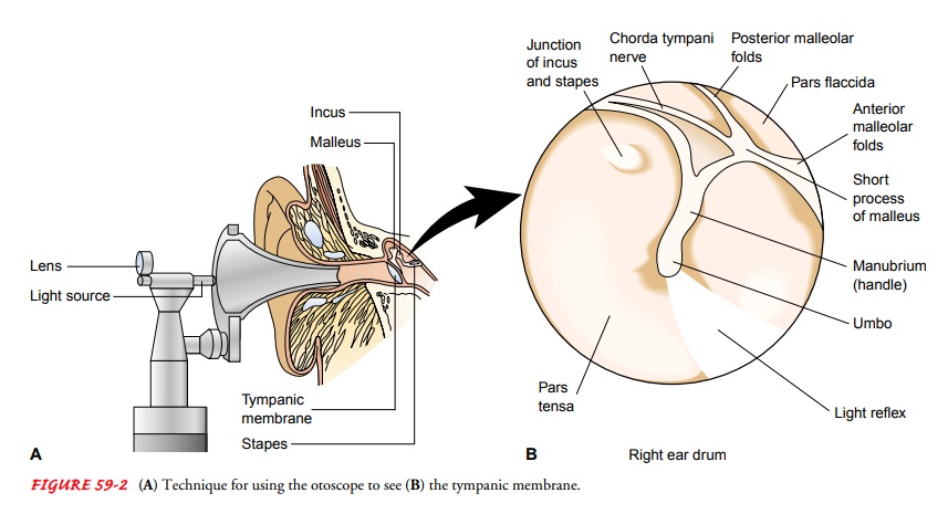
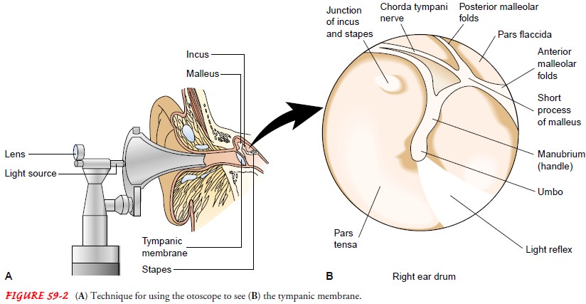
Tympanic Membrane
The tympanic membrane (ie, eardrum), about 1 cm in diame-ter and very thin, is normally pearly gray and translucent. The tympanic membrane consists of three layers of tissue: an outer layer, continuous with the skin of the ear canal; a fibrous mid-dle layer; and an inner mucosal layer, continuous with the lining of the middle ear cavity. Approximately 80% of the tympanic membrane is composed of all three layers and is called the parstensa.
The other 20% of the tympanic membrane lacks the middle layer and is called the
pars flaccida. The absence of this fibrous middle layer makes the pars flaccida
more vulnerable to pathologic disorders than the pars tensa. Distinguishing
land-marks of the tympanic membrane include the annulus, the fi-brous border
that attaches the eardrum to the temporal bone; the short process of the
malleus; the long process of the mal-leus; the umbo of the malleus, which
attaches to the tympanic membrane in the center; the pars flaccida; and the
pars tensa (Fig. 59-2).
The
tympanic membrane protects the middle ear and con-ducts sound vibrations from
the external canal to the ossicles. The sound pressure is magnified 22 times as
a result of transmission from a larger area to a smaller one.
Ossicles
The
middle ear contains the three smallest bones (ie, ossicles) of the body:
malleus, incus, and stapes. The ossicles, which are held in place by joints,
muscles, and ligaments, assist in the transmis-sion of sound. Two small
fenestrae (ie, oval and round windows), located in the medial wall of the
middle ear, separate the middle ear from the inner ear. The footplate of the stapes
sits in the oval window, secured by a fibrous annulus, or ring-shaped
structure. The footplate transmits sound to the inner ear. The round win-dow,
covered by a thin membrane, provides an exit for sound vibrations (see Fig.
59-1).
Related Topics