Chapter: Medical Surgical Nursing: Assessment of Digestive and Gastrointestinal Function
Anatomy of the Gastrointestinal Tract
ANATOMY
OF THE GASTROINTESTINAL TRACT
The GI
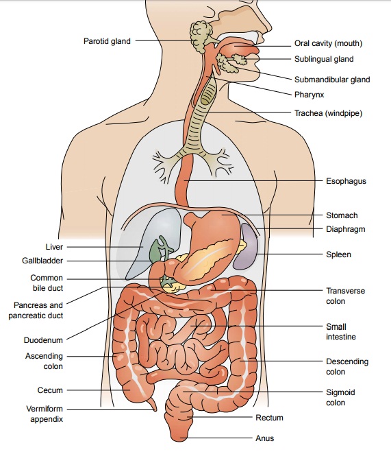
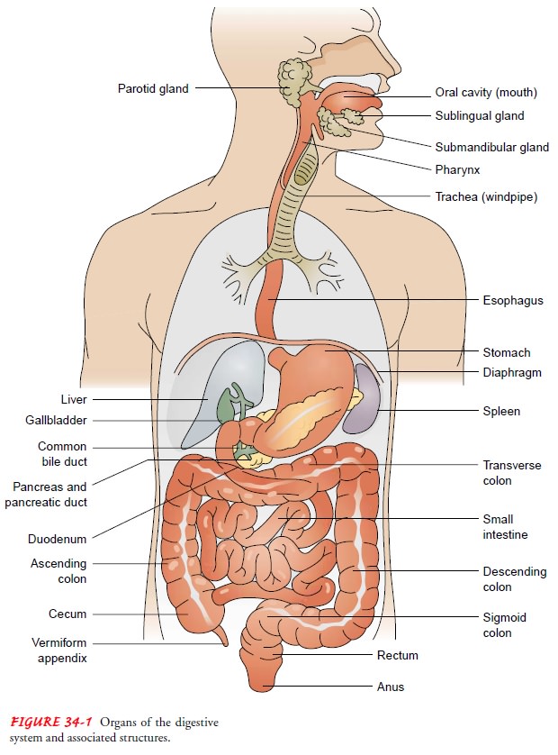
tract is a 23- to 26-foot-long pathway that extends from the mouth through the esophagus, stomach,
and intestines to the anus (Fig. 34-1). The esophagus is located in the mediastinum in the thoracic cavity,
anterior to the spine and posterior to the trachea and heart. This collapsible
tube, which is about 25 cm (10 inches) in length, becomes distended when food
passes through it. It passes through the diaphragm at an opening called the
diaphragmatic hiatus.
The
remaining portion of the GI tract is located within the peritoneal cavity. The stomach is situated in the upper
portion of the abdomen to the left of the midline, just under the left
di-aphragm. It is a distensible pouch with a capacity of approximately 1500 mL.
The inlet to the stomach is called the esophagogastric junction; it is
surrounded by a ring of smooth muscle called the lower esophageal sphincter (or
cardiac sphincter), which, on con-traction, closes off the stomach from the esophagus.
The stomach can be divided into four anatomic regions: the cardia (entrance),
fundus, body, and pylorus (outlet). Circular smooth muscle in the wall of the
pylorus forms the pyloric sphincter and controls the opening between the
stomach and the small intestine.
The small intestine is the longest segment
of the GI tract, accounting for about two thirds of the total length. It folds
back and forth on itself, providing approximately 7000 cm of sur-face area for
secretion and absorption, the
process by which nu-trients enter the bloodstream through the intestinal walls.
The small intestine is divided into three anatomic parts: the upper part,
called the duodenum; the middle part, called the jejunum; and the lower part,
called the ileum. The common bile duct, which allows for the passage of both
bile and pancreatic secre-tions, empties into the duodenum at the ampulla of
Vater. The junction between the small and large intestine, the cecum, is
lo-cated in the right lower portion of the abdomen. The ileocecal valve is
located at this junction. It controls the passage of intesti-nal contents into
the large intestine and prevents reflux of bacte ria into the small intestine.
The vermiform appendix is located near this junction.
The large intestine consists of an
ascending segment on the right side of the abdomen, a transverse segment that
extends from right to left in the upper abdomen, and a descending segment on
the left side of the abdomen. The terminal portion of the large in-testine
consists of two parts: the sigmoid colon and the rectum. The rectum is
continuous with the anus. A network
of striated mus-cle that forms both the internal and the external anal
sphincters regulates the anal outlet.
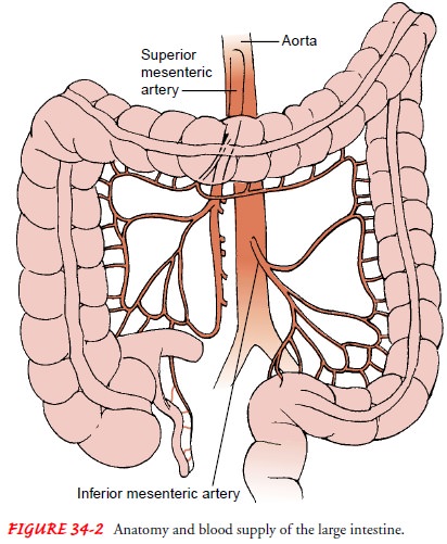
The GI
tract receives blood from arteries that originate along the entire length of
the thoracic and abdominal aorta. Of partic-ular importance are the gastric
artery and the superior and infe-rior mesenteric arteries. Oxygen and nutrients
are supplied to the stomach by the gastric artery and to the intestine by the mesen-teric
arteries (Fig. 34-2). Blood is drained from these organs by veins that merge
with others in the abdomen to form a large ves-sel called the portal vein.
Nutrient-rich blood is then carried to the liver. The blood flow to the GI
tract is about 20% of the total cardiac output and increases significantly
after eating.
Both
the sympathetic and parasympathetic portions of the auto-nomic nervous system
innervate the GI tract. In general, sympa-thetic nerves exert an inhibitory
effect on the GI tract, decreasing gastric secretion and motility and causing
the sphincters and blood vessels to constrict. Parasympathetic nerve
stimulation causes peri-stalsis and increases secretory activities. The
sphincters relax under the influence of parasympathetic stimulation. The only
portions of the tract that are under voluntary control are the upper esophagus
and the external anal sphincter.
Related Topics