Chapter: Medical Surgical Nursing: Assessment and Management of Female Physiologic Processes
Anatomy of the Female Reproductive System
ANATOMY
OF THE FEMALE REPRODUCTIVE SYSTEM
The
female reproductive system consists of external and internal structures. Other
anatomic structures that affect the female re-productive system include the
hypothalamus and pituitary gland of the endocrine system.
External Genitalia
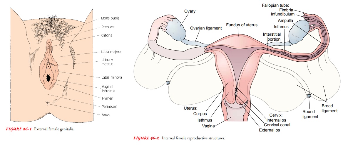
The external genitalia (the vulva) include two thick folds of tissue called the labia majora and two smaller lips of delicate tissue called the labia minora, which lie within the labia majora. The upper por-tions of the labia minora unite, forming a partial covering for the clitoris, a highly sensitive organ composed of erectile tissue. Be-tween the labia minora, below and posterior to the clitoris, is the urinary meatus. This is the external opening of the female urethra and is about 3 cm (1.5 inches) long. Below this orifice is a larger opening, the vaginal orifice or introitus (Fig. 46-1). On each side of the vaginal orifice is a vestibular (Bartholin’s) gland, a bean-sized structure that empties its mucous secretion through a small duct. The opening of the duct lies within the labia minora, external to the hymen. The area between the vagina and rectum is called the perineum.
Internal Reproductive Structures
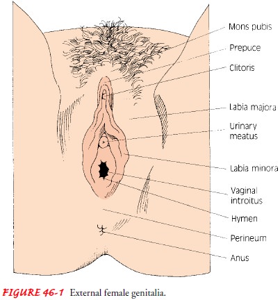
The
internal structures consist of the vagina, uterus, ovaries, and fallopian or
uterine tubes (Fig. 46-2).
VAGINA
The
vagina, a canal lined with mucous membrane, is 7.5 to 10 cm (3 to 4 inches)
long and extends upward and backward from the vulva to the cervix. Anterior to
it are the bladder and the urethra, and posterior to it lies the rectum. The anterior
and posterior walls of the vagina normally touch each other. The upper part of
the vagina, the fornix, surrounds
the cervix (the inferior part of the
uterus).
UTERUS
The
uterus, a pear-shaped muscular organ, is about 7.5 cm (3 inches) long and 5 cm
(2 inches) wide at its upper part. Its walls are about 1.25 cm (0.5 inch)
thick. The size of the uterus varies, depending on parity (number of viable
births) and uterine abnormalities (eg, fibroids, which are a type of tumor that
may distort the uterus). A nulliparous woman (one who has not com-pleted a
pregnancy to the stage of fetal viability) usually has a smaller uterus than a
multiparous woman (one who has com-pleted two or more pregnancies to the stage
of fetal viability). The uterus lies posterior to the bladder and is held in
position by sev-eral ligaments. The round ligaments extend anteriorly and
later-ally to the internal inguinal ring and down the inguinal canal, where
they blend with the tissues of the labia majora. The broad ligaments are folds
of peritoneum extending from the lateral pelvic walls and enveloping the
fallopian tubes. The uterosacral ligaments extend posteriorly to the sacrum.
The uterus has two parts: the cervix, which projects into the vagina, and a
larger upper part, the fundus or
body, which is covered posteriorly and partly anteriorly by peritoneum. The
triangular inner portion of the fundus narrows to a small canal in the cervix
that has con-strictions at each end, referred to as the external os and
internal os. The upper lateral parts of the uterus are called the cornua. From
here, the oviducts or fallopian (or uterine) tubes extend outward, and their
lumina are internally continuous with the uterine cavity.
OVARIES
The ovaries lie behind the broad ligaments,
behind and below the fallopian tubes. They are oval bodies about 3 cm (1.2
inches) long. At birth, they contain thousands of tiny egg cells, or ova. The
ovaries and the fallopian tubes together are referred to as the adnexa.
Related Topics