Chapter: Essentials of Anatomy and Physiology: Digestive System
Anatomy and Histology of the Digestive System
ANATOMY AND HISTOLOGY OF THE DIGESTIVE SYSTEM
The digestive system consists of the digestive tract, or gastro-intestinal (GI; gas′trō-in-tes′tin-ăl) tract, plus specific associatedorgans. Because the digestive tract is open at the mouth and anus, the inside of the tract is continuous with the outside environment, and food entering the digestive tract may contain not only useful nutrients but also indigestible components such as fiber, or harmful materials such as bacteria. Therefore, the inner lining of the diges-tive tract serves as a protective barrier to those indigestible and harmful materials and nutrients must be specifically transported across the wall of the digestive tract. Once across the wall of the digestive tract, the nutrients enter the circulation to access tissues of the body.
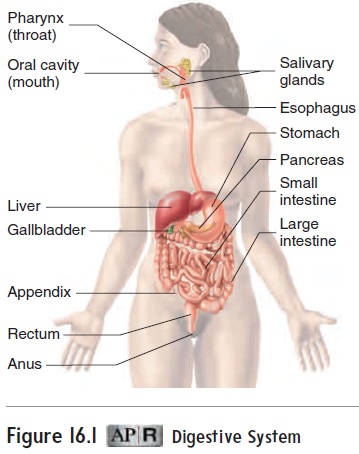
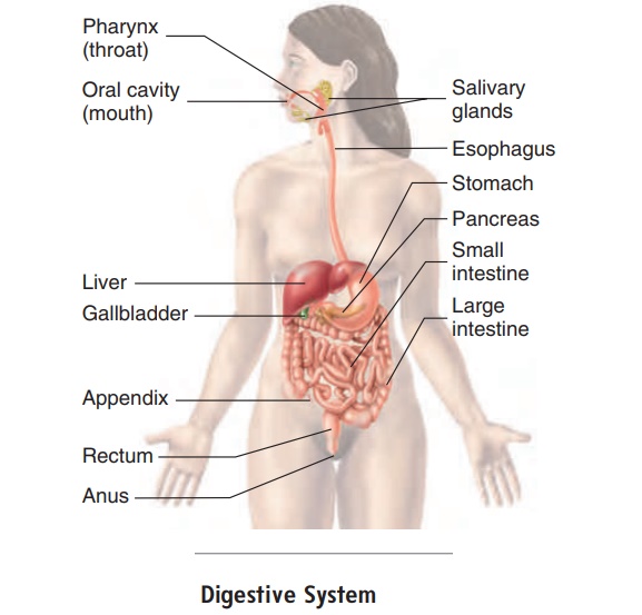
The digestive tract consists of the oral cavity, pharynx, esophagus, stomach, small intestine, large intestine, and anus. Accessory glands are associated with the digestive tract (fig-ure 16.1). The salivary glands empty into the oral cavity, and the liver and pancreas are connected to the small intestine.
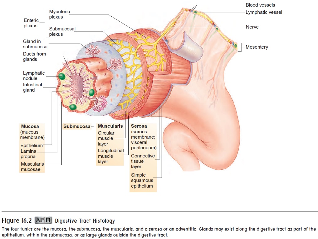
Various parts of the digestive tract are specialized for differ-ent functions. Nearly all segments of the digestive tract consist of four layers, called tunics. These are the mucosa, the submucosa, the muscularis, and a serosa or an adventitia (figure 16.2):
1. The innermost tunic, the mucosa (mū-kō′ să), consists of mucous epithelium, a loose connective tissue called the lamina propria, and a thin smooth muscle layer, the muscularis mucosae. The epithelium in the mouth,esophagus, and anus resists abrasion, and the epithelium in the stomach and intestine absorbs and secretes.
2. The submucosa lies just outside the mucosa. It is a thick layer of loose connective tissue containing nerves, bloodvessels, and small glands. An extensive network of nerve cell processes forms a plexus(network). Autonomic nerves innervate this plexus.
3. The next tunic is the muscularis. In most parts of the digestive tract it consists of an inner layer of circularsmooth muscle and an outer layer of longitudinal smooth muscle. Another nerve plexus, also innervatedby autonomic nerves, lies between the two muscle layers.Together, the nerve plexuses of the submucosa and muscularis compose the enteric (en-tĕr′ ik) nervoussystem. This nervous system, which is a division of theautonomic nervous system, is extremely important in controlling movement and secretion within the tract .
4. The fourth, or outermost, layer of the digestive tract is either a serosa or an adventitia. The serosa consists of the peritoneum, which is a smooth epithelial layer, and its underlying connective tissue. Regions of the digestive tractnot covered by peritoneum are covered by a connective tissue layer called the adventitia (ad′ ven-tish′ ă; foreign, coming from outside), which is continuous with the surrounding connective tissue.
Peritoneum
The body wall of the abdominal cavity and the abdominal organs is covered with serous membranes (figure 16.3). The serous mem-brane that covers the organs is the serosa, or visceral peritoneum (per′ i-tō-nē′ ŭm; to stretch over). The serous membrane that lines the wall of the abdominal cavity is the parietal peritoneum.
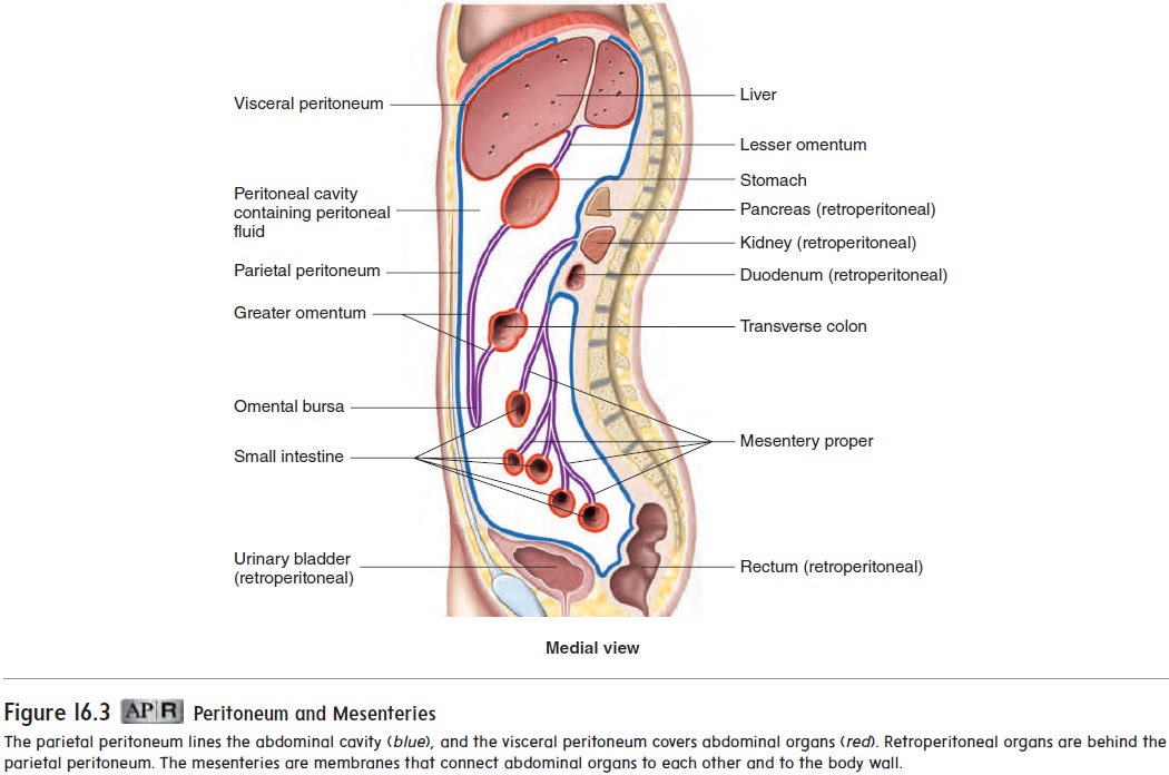
Many of the organs of the abdominal cavity are held in place by connective tissue sheets called mesenteries (mes′ en-ter-ēz). Mesentery is a general term referring to the serous membranesattached to the abdominal organs. The mesenteries consist of two layers of serous membranes with a thin layer of loose connective tissue between them. The mesentery connecting the lesser curva-ture of the stomach to the liver and diaphragm is called the lesseromentum (ō-men′tŭm), and the mesentery connecting the greatercurvature of the stomach to the transverse colon and posterior body wall is called the greater omentum. The greater omentum is unusual in that it is a long, double fold of mesentery that extends inferiorly from the stomach before looping back to the transverse colon to create a cavity, or pocket, called the omental bursa (ber′ să). Adipose tissue accumulates in the greater omentum, giving it the appearance of a fat-filled apron that covers the anterior surface of the abdominal viscera. The mesentery that attaches the small intestine to the posterior abdominal wall is called the mesentery proper.
Other abdominal organs lie against the abdominal wall, have no mesenteries, and are described as retroperitoneal (re′ trō-per′ i-tō-nē′ ăl; behind the peritoneum). The retroperitoneal organs include the duodenum, pancreas, ascending colon, descending colon, rectum, kidneys, adrenal glands, and urinary bladder.
Related Topics