Chapter: Medical Physiology: Electrocardiographic Interpretation of Cardiac Muscle and Coronary Blood Flow Abnormalities: Vectorial Analysis
Abnormalities in the T Wave - Electrocardiographic Interpretation of Cardiac Muscle and Coronary Blood Flow Abnormalities
Abnormalities in the T Wave
Earlier, it was pointed out that the T wave is normally positive in all the standard bipolar limb leads and that this is caused by repolarization of the apex and outer surfaces of the ventricles ahead of the intraventricular surfaces. That is, the T wave becomes abnormal when the normal sequence of repolarization does not occur. Several factors can change this sequence of repolarization.
Effect of Slow Conduction of the Depolarization Wave on the Characteristics of the T Wave
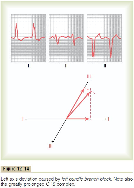
Referring back to Figure 12–14, note that the QRS complex is considerably prolonged. The reason for this prolongation is delayed conduction in the left ventricle resulting from left bundle branch block. This causes the left ventricle to become depolarized about 0.08 second after depolarization of the right ventricle, which gives a strong mean QRS vector to the left. However, the refractory periods of the right and left ventricular muscle masses are not greatly different from each other. Therefore, the right ventricle begins to repolarize long before the left ventricle; this causes strong positivity in the right ventricle and negativity in the left ventricle at the time that the T wave is devel-oping. In other words, the mean axis of the T wave is now deviated to the right, which is opposite the mean electrical axis of the QRS complex in the same elec-trocardiogram. Thus, when conduction of the depolar-ization impulse through the ventricles is greatly delayed, the T wave is almost always of opposite polar-ity to that of the QRS complex.
Shortened Depolarization in Portions of the Ventricular Muscle as a Cause of T Wave Abnormalities
If the base of the ventricles should exhibit an abnor-mally short period of depolarization, that is, a short-ened action potential, repolarization of the ventricles would not begin at the apex as it normally does.
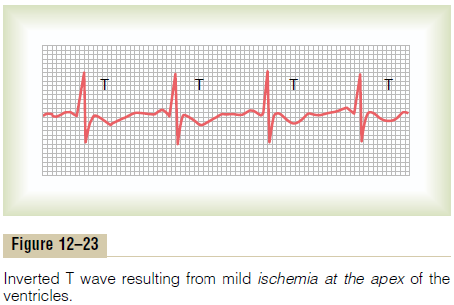
Instead, the base of the ventricles would repolarize ahead of the apex, and the vector of repolarization would point from the apex toward the base of the heart, opposite to the standard vector of repolariza-tion. Consequently, the T wave in all three standard leads would be negative rather than the usual positive. Thus, the simple fact that the base of the ventricles has a shortened period of depolarization is sufficient to cause marked changes in the T wave, even to the extent of changing the entire T wave polarity, as shown in Figure 12–23.
Mild ischemia is by far the most common cause ofshortening of depolarization of cardiac muscle, because this increases current flow through the potas-sium channels. When the ischemia occurs in only one area of the heart, the depolarization period of this area decreases out of proportion to that in other portions. As a result, definite changes in the T wave can take place. The ischemia might result from chronic, pro-gressive coronary occlusion; acute coronary occlusion; or relative coronary insufficiency that occurs during exercise.
One means for detecting mild coronary insufficiency is to have the patient exercise and to record the elec-trocardiogram, noting whether changes occur in the T waves. The changes in the T waves need not be spe-cific, because any change in the T wave in any lead— inversion, for instance, or a biphasic wave—is often evidence enough that some portion of the ventricular muscle has a period of depolarization out of propor-tion to the rest of the heart, caused by mild to moder-ate coronary insufficiency.
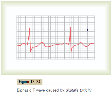
Effect of Digitalis on the T Wave. As discussed, digitalis is a drug that can be used during coronary insufficiency to increase the strength of cardiac muscle contraction. But when overdosages of digitalis are given, depolarization duration in one part of the ven-tricles may be increased out of proportion to that of other parts. As a result, nonspecific changes, such as T wave inversion or biphasic T waves, may occur in one or more of the electrocardiographic leads. A biphasic T wave caused by excessive administration of digitalis is shown in Figure 12–24. Therefore, changes in the T wave during digitalis administration are often the ear-liest signs of digitalis toxicity.
Related Topics